Categories
— Concept
— Chapter 1: The Body as Object of Study: Anatomical Diagrams from the 12th–13th Centuries
— Chapter 2: The Body as System: Depiction of Internal Organs and Structures
— Chapter 3: The Body as Guide for Treatment: Medical Schematics from the 15th Century
— Bibliography
— Image Sources
Concept
In the 12th–15th centuries, depictions of the human body played a crucial role in conveying medical knowledge. They served as a form of visual aid for physicians and those studying medicine, allowing them to represent the anatomical features of the body within the knowledge available during the Middle Ages. Ancient traditions, Galen’s teachings, and astrological beliefs influenced medical graphics, determining the methods of illustrating and interpreting the human form. This period was marked not only by significant artistic achievements but also by epidemics, wars, and the spread of superstitions. Against this backdrop, the development of medical knowledge and methods of its visual transmission took on special significance. Medieval medical graphics are interesting precisely because they combine artistic expression with practical function.
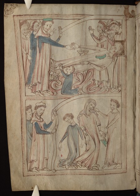
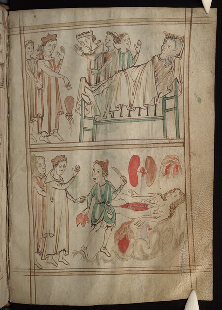
Ashmole Manuscript 399, late 13th — early 14th century.
The objective of this study is to trace the evolution of how the human body was visually depicted in medical graphics from the 12th to the 15th centuries—from anatomical diagrams and illustrations of internal organs to depictions of medical procedures. Special attention is paid to the specifics of body representation, the use of conventional forms, and the conveyance of medical concepts through graphic media.
Chapter 1: The Body as an Object of Study: Anatomical Diagrams from the 12th–13th Centuries
In medieval medicine, the ancient heritage played a vital role, particularly the works of the ancient Greek physician Galen. His views on the structure of the human body remained authoritative for centuries and were used in medical education. However, many of Galen’s anatomical understandings were based on the dissection of animals—primarily monkeys and pigs—which led to certain inaccuracies in describing the human body.
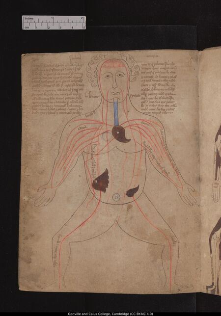
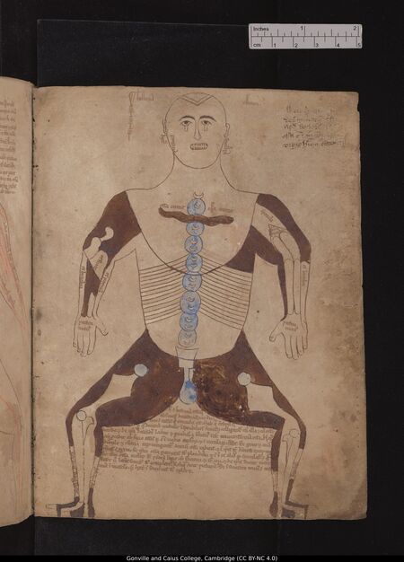
An anatomical series of nine figures, circa 1200, Gonville and Caius College, MS 190/223
These representations are also reflected in medieval medical graphics. Anatomical illustrations did not aim for a realistic depiction of the human body. Their purpose was to clearly present individual organ systems and make complex information accessible for study. This chapter examines anatomical series from the 12th–13th centuries. The main focus is on how the inner structures of the human body were depicted using the figure’s positioning, lines, color, and captions.
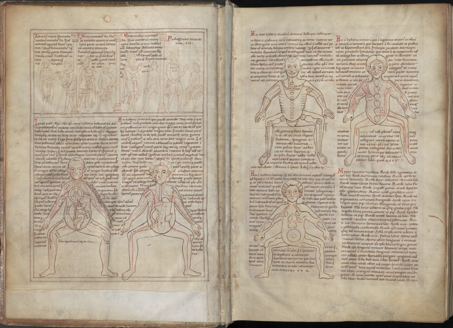
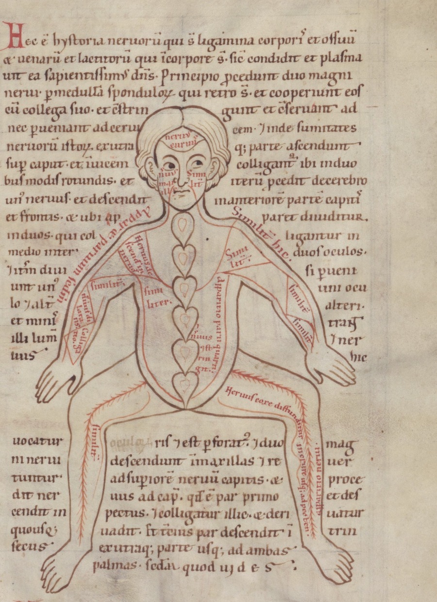
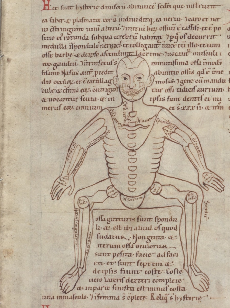
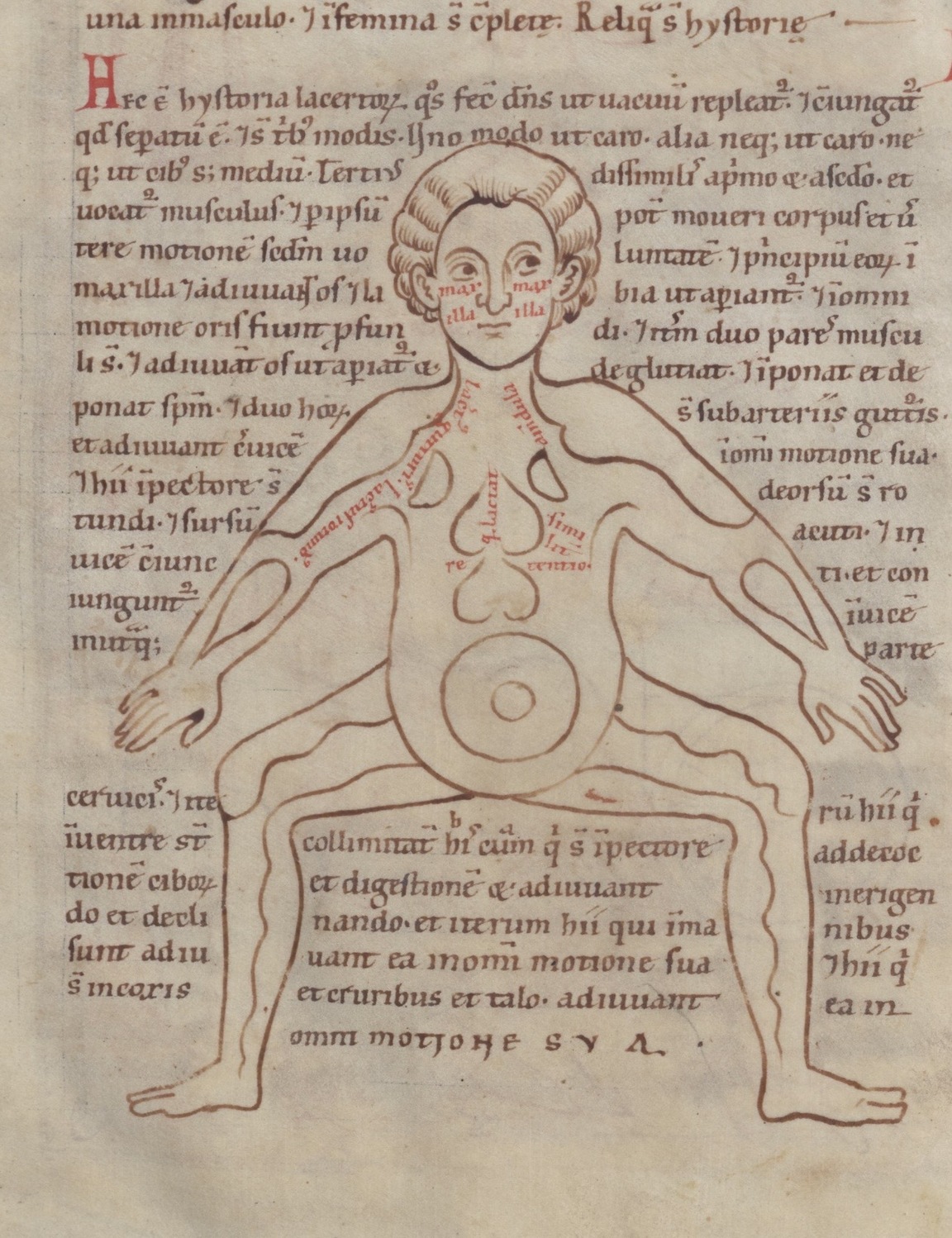
Anatomical series of five figures, circa 1165, Clm 13002, folios 2v–3r
The first example is the «Anatomical Series of Five Figures» (mid-12th century), presented in the manuscript Clm 13002. It comprises five depictions of squatting human figures, each illustrating a separate body system: veins, arteries, bones, nerves, and muscles. The series draws upon the ancient anatomical tradition and is linked to Galenic understandings of bodily structure. The recurring depiction of the human figure allows for the examination of each anatomical system in isolation. Despite this, the overall silhouette remains recognizable while its internal components change. Through this technique, the viewer can focus on a specific structure without being distracted by other elements of the body.
The figure’s pose is particularly notable: the person is depicted squatting, with bent and spread legs. This posture creates a compact yet sufficiently broad silhouette, within which the branching lines of blood vessels, nerves, and other anatomical elements can be placed. The body proportions are also subservient to the representational goal. Individual parts of the figure appear enlarged or simplified compared to the actual human body. This allows for the inclusion of more detail and makes the internal structure more visible.